2007-May-21 (月) 22:53 +8:00
☆ 季節感なし・No Seasonal Feeling
The temperature is turning warm from May. Though Beginnig of Summer was 6th May this year, in common, it should be June when we put on our summer clothing. And in the late period of May, the weather has turned very hot too. It is really like summer now.
So that, the spring and summer in this year had became a mess. For example, morning glories, which should come in July, but I have seen them in the beginning of May; water lilies, which should come in August, now they have bloomed in my campus; and dandelions, which should come in March, can be seen even now the late May.
I was surprised completely for all of them. What the season is now on earth?
Do not you feel puzzled about it?
All of us might be pulled in fact, I think.
Living in the modern, I still not want to forget the tradition. Like the seasonal feelings.
Surely, I most like summer and autumn, and I also not dislike the others. Thanks for them, I can enjoy my like seasons more strongly and cherished. Do not you think so?
Nevertheless, it is a little regret. Climate started to turning warm before many years. Therefore, the seasonal feeling also start vanished from us.
The seasonal feeling is important to me. Flowers, rains, leaves, and snows, each season has its own unique beauties. And with the changing seasons, he beauties transformed silently, tenuously and lightly. The former and the later season overlaps each other, one went and the next come. Sometimes, as I find out any tiny change suddenly, a happy also cherished feeling come into my heart. This tender mood is must a present from the nature, and, it is must a big appreciation which respond to the mysterious nature from me.
Now, seeing everyone and me in the summer clothing, somewhat, I think a bit about the seasonal feeling will be vanished soon.
皐月になってから、気温はますます暑くなっています。5月6日は立夏というものですけど、普通、半袖なんてを着るのは6月のことはずです。でも、この皐月の中旬に、私たちはもう半袖を着ることになりました。そして、下旬に入ってきた「今」は、気温はとても暑くなってしまいました。まるでほんものの夏みたいです。
という訳で、今年の春と夏もむちゃむちゃになってきました。例えば、もともと、文月の季語「朝顔」はね、皐月の初めにも見られることになりました;また、葉月の季語「蓮」も、私のキャンパスに満開してきました;それにもう一つ、弥生の季語「蒲公英」、今、この皐月の下旬までも見られます。
まったく驚かれました。今、いったい何の季節なんですか?
あなたも、こんなことに本当に迷うことなんでしょう?
私、現代に生きているけど、伝統的なものを捨てたくないんです。その季節感も、この一つです。
一年中、私の一番好きな季節が夏と秋はもちろん、でもほかの季節にも嫌いことではありません。ほかの季節があるこそ、好きな季節がより愛しく楽しんでいられることができますから。そうじゃありませんか?
けど、残念ながら、何年か前から、気候は暑くなり始めました。季節感もだんだん私のそばから消え続けています。
季節感は私にとって大切なものです。花雨葉雪、一つ一つの季節にも、その季節なりのつくどくな美しさが持っています。また、この美しさは、それぞれ季節の移り回ると共に、静寂で繊細で微妙で小さく変わっています。この季節と次の季節が、お互いに重ねて消えちゃって、そして生まれて。時折、不意に何かの変化を見つめると、心も楽しかった惜しんだりします。この愛しい心地はきっと、自然からもらった贈り物です、これもきっと、自然の不思議さへの大きいご感謝です。
今、半袖の皆さんと私を見ていて、何とかちょっぴり、季節感が本当に消えちゃうような気がします。
2007-May-19 (土) 24:53 +8:00
♤ 粉嗜好症・Powder-Mania
At first, look at the photo please.
始めに、この写真を見てください
始めに、この写真を見てください
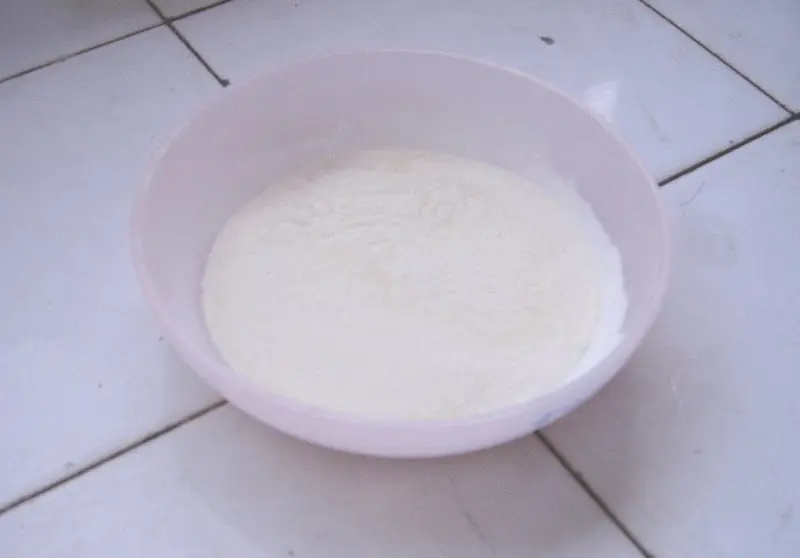
Do you know what it is?
これは何だと、お存じますか?
これは何だと、お存じますか?
Now, it’s the answer.
That\'s the powder of a soap.
はい、これは答えです。
石鹸の粉なのです。
That\'s the powder of a soap.
はい、これは答えです。
石鹸の粉なのです。
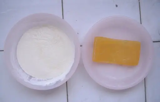
Don\'t you surprise? Well, there is something more surprise. So I need say some words about the powder itself.
Please think about it: you, with your right hand a piece of metal, and with your left hand a tiny part of soap. Then let\'s start.
Softly, you scrape the soap with your metal piece on the very surface of the soap, so that the fine powder fall into the readied tray. Do it continuing, the powder will be piled, so you can see the soft, fine and light powder of a soap.
It\'s a trouble work sure enough, common people, like me, may have no any patience about it. However, this kind who can do it is here beside me.
Mio-chan, she made the powder. As my know, everyday, she did little on it, and I could get this photos finally.
In her own words, she was born with this affinity on powder. It\'s really, certainly she love all the powders. Therefore, I call her "the Powder-Mania".
But, what a strange hobby she has!
So the powder which she had a hard time to make, could be effective in any washing?
驚いたんでしょう?でも、まだより驚かせることがありますよ。この粉自身について、私、一言があります。
ちょっとご想像してください、あなたの右手に、ある小さな金属片を持っていて、左手に、一つすぐに使いきれる石鹸を持っています。そして、あなたは始めました。
その金属片で石鹸の一番表面から軽く滑り覆って、この間、設計の細かい粉がゆらゆらと、用意してきた器の中に落ちました。こうして続いたら、器に落ちた粉もだんだん積もりだした。それで、この柔らかで細かくてふわふわな石鹸の粉が、あなたの目の前に浮かべだした。
本当になかなか大変ですね。私など普通の人は、こんな面倒くさいことに、あまり耐えらなくなるに決まっています。でも、こんなことを上手に出来上がれる人は、すぐ私の傍にいます。
未央ちゃん、この粉は彼女が出来上がったものです。私の知っている限り、彼女は毎日ずつ、少しだけを作っていて、そして、私はこの写真ができました。
彼女自身の言葉で言えば、彼女の粉に強くてならない愛着は、生まれながらのものです。確かに、彼女はすべての粉に強く愛していますの。ですから、「粉嗜好症」私はこれで彼女を呼べます。
やはり変な癖です…
で、やっと出来上がったこの粉は、まだ洗うことに役立ていますか?
2007-May-04 (金) 13:06 +8:00
♡ この想い、捨てられないで・The Feeling I Can not Throw Away
I had lots of classmates when I was in my high school, and now, few are keep touching. And in such a time, I realized what's the real friendship.
The common communication is too superficial to me, but I must be very joyful if someone did something for me at the time which important to me. This feeling must more joyful and must be millions times than any others. It need not extraordinary, though little things such as a comfort, a timely greeting, a saying "I' back". It can be anything. And after the short meeting time ended, I'm on my own again, however, suddenly, my heart have filled with the endless happiness.
"For we are friends, aren’t we?"
Although it's such a simple sentence, it can make me reach the blessing deeply.
If it is not a magic, what it is?
Surely I know the magic of friendship long ago, but the time when I really perceive it, was last night.
She, Misato-chan, was one of my friends in my high school. When we were in 3 grade, she immigrated to Canada. Hence we were rarely linked beside some emails. And with the time elapsing, she is fading in my heart.
The night before yesterday, a message from Yan-kun told me that she was back, in that moment, all about her and me flowed out from my heart.
I remembered clearly the day of her birthday 3 years ago. Ichika-chan and me gave her our hand-made present, and soon we got her reply. Now that reply is put in my treasure box in my room. Each time, when I unfold the paper and read it, I feel… how nostalgic!
After so long time we did not touch, in the so-called "now", I can still remember all the thing in the past time. That's must be the magic of friendship certainly.
Now, she was back, it would be a reunion after the 3 years. When I knew the news then, my heart excited. But I couldn't attend the party but had to stay in college. So I sent my fact and regret. But I didn't know why I still couldn't turn to calms after that.
I realized there was something lacked.
I want attend too, in fact.
Though I am not good at communication, but then, I had a kind of mood that I wanted to go no matter what happened.
At last, I knew I couldn't, because of the fact I was not at home but at college.
Thus the next day came.
I realized there was something lacked.
I want attend too, in fact.
Though I am not good at communication, but then, I had a kind of mood that I wanted to go no matter what happened.
At last, I knew I couldn't, because of the fact I was not at home but at college.
Thus the next day came.
The party would be started at 18 o'clock, and till 22 o'clock, I past peacefully, besides, my kind of void heart, why?
About 10:10, Ichika-chan told me, their party would be ended.
I found out why I felt void from this evening.
Originally I wanted to meet Misato-chan in this summer holiday, but also Ichika-chan, told me that she would back to Canada few days after, and from that would be another 3 years.
It was likely everything freezed.
If I still like this… no, I must do something, I had not any reason to do nothing.
No matter it was a long-distance phone, I wanted to speak to her in the night!
So, later, I called her,
So, later, her answered,
So, later, we talked happily,
Like before.
And, later,
"Now" came, and I smiled satisfyingly.
That was surely very regretful for I couldn't attend the party, but my heart was turning not so void.
It was so good for I called her.
Nowadays, the more I tough my old friends, I feel so more:
Remember them all the time,
And, don't forget this feeling.
Remember them all the time,
And, don't forget this feeling.
高校生の頃、たくさんの仲間があった。今まで、もう何人しかそのまま連絡していないの。そして、こんな時には、本当の友達なんてを、やっと気付いてきたの。
普通の付き合いはただ表面的の浅くてならないこと、でも、自分にとって大切な時に、友から何かがしてくれたら、普通の時より、何千何万倍で喜びになれることに決まっているの。それがどのくらいたいした事じゃなくても、それがたった一つ小さなことだけでも、たとえ一つの慰め、一つ小さな時より挨拶、或いは「ただいま」って言葉。何でも、何でも。そして、短い付き合いが終わった時、自分はまだ一人に戻った。けど、心がふっと幸せと感心な気持ちに満ちられた。
「友達だから、ねえ。」
こんな簡単な言葉だけど、心にこんな深く深く喜ばせることができるの。
これは魔法じゃなったら、何だろう?
友情の不思議なんて、よく分かったけど、私は本当にこれを感じることは、昨夜は初めてだった。
彼女、美里ちゃんは、高校生の友達だった、3年生の頃、カナダに移動してきた。それから、私たちは偶にいくつのメールのほか、ぜんぜん連絡しなかった。時間の移りと共に、少しずつ少しずつ、そのこのことは私の心に消えちゃっていた。
でも、一昨日の夜、やんくんからメッセージがもらった、彼女が戻ってきたと教えてくれた。
一瞬間、その高校生の3年間、彼女と一緒に過ごしたすべてが、心から溢れ出した。
まだ覚えているの、3年前彼女の誕生日に。私といちかちゃんは彼女に手作りのプレゼントを贈った。そしてすぐに彼女から返事をもらった。その返事、今も私の部屋の宝物の小箱においている。その小さな紙を開けて読むたびに、懐かしくて懐かしくてならないの。
長い間連絡なかったけど、この「今」でも、その時のすべてをも思い出せるのが、きっと友情の不思議な魔法だね。
でも、一昨日の夜、やんくんからメッセージがもらった、彼女が戻ってきたと教えてくれた。
一瞬間、その高校生の3年間、彼女と一緒に過ごしたすべてが、心から溢れ出した。
まだ覚えているの、3年前彼女の誕生日に。私といちかちゃんは彼女に手作りのプレゼントを贈った。そしてすぐに彼女から返事をもらった。その返事、今も私の部屋の宝物の小箱においている。その小さな紙を開けて読むたびに、懐かしくて懐かしくてならないの。
長い間連絡なかったけど、この「今」でも、その時のすべてをも思い出せるのが、きっと友情の不思議な魔法だね。
彼女が戻ってきたの。これが3年ぶりの再会だよ。その一瞬、本当に心がわくわくした。でも、私は学校に渋滞していて戻れなかったの。しようがなかったね、パーティーに出られないと自分の残念な心情をみんなに伝ったけど、どうしてなのか、心がずっと平気でいられなかったの。
何かが済まないように気がした。
私も行きたかったのよ、実は。
ほかの人に交流することなんて、あまり上手じゃないけど、これだけ、美里ちゃんだから、どうしても出て行きたい…という心情だった。
しかし、やはり無理だったよね、学校に渋滞した私が…
そして、次の日がやって来た。
何かが済まないように気がした。
私も行きたかったのよ、実は。
ほかの人に交流することなんて、あまり上手じゃないけど、これだけ、美里ちゃんだから、どうしても出て行きたい…という心情だった。
しかし、やはり無理だったよね、学校に渋滞した私が…
そして、次の日がやって来た。
パーティーの時間は、午後6時だった。夜10時まで私も平気ですぎった。ただ、心が空っぽかったの。どうしてね?
10時10分頃、いちかちゃんから、パーティーはもうすぐ終わりだよと知らせてくれた。
やっと気づいた、私の空っぽくなった訳を。
今は戻れないから、もともと、夏休みに美里ちゃんと会おうと想ったけど、彼女はいくつか日のあとでカナダに帰るということも、いちかちゃんから分からせた。
全てが凍えられたようだった。
このままじゃ…もうだめ、私、何とかしなきゃ!もう待っている訳には行けないの!
長距離電話でもいいの。今夜こそ、きっと話し合いたいの!
そのあとは、電話した、
そのあとは、彼女が答えた、
そのあとは、二人は楽しく話し合った、
昔のようにだったの。
そのあとは、「今」がやって来た、私は済んで笑っていた。
パーティーに出られなかった、ちょっぴり残念だった。でも、私の心はもうそんなに空っぽくなくなるんだ。
電話してあげて、よかった。
今、昔の友達と繋げれば繋ぐほど、私はこんな感じがするの。
この気持ちを永遠に覚えておいで、
そして、この想い、捨てられないで。
この気持ちを永遠に覚えておいで、
そして、この想い、捨てられないで。
2007-May-02 (水) 15:05 +8:00
☆ 野イチゴの地・Here the Wild Strawberries
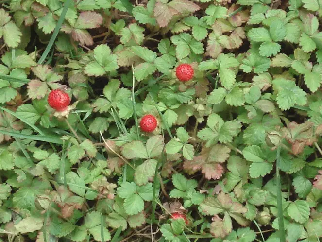
There are lots of wild strawberries in my campus.
I don't know whether they are wild strawberries, but I call them only as my classmates so told me.
And finally when I wanted to take photos for them a few days before, I have seen them long time from the beginning of April.
The wild strawberries were red and little, to pick one, the fruit was hard. If you rub the fruit, the littler red seeds fallen granule by granule. Besides, there was also faint scent drifted out.
Stand in a farther place to see them. On the green lawn, the little wild strawberries were growing at all places, dazzling in the sunshine. What a comfortable and lovely scene it was!
キャンパスのあるところ、たくさんの野イチゴがあります。
それが野イチゴかどうか、私はよく分かりませんが、クラスメートたちもそう言っているから、一応、野イチゴに見知れましょう。
四月にこれを見てきましたが、先日まで、写真撮る欲望が浮かべだした。
一つ一つの野イチゴは真っ赤で、小さくて。一粒を摘み見ると、実りが硬く、小さめの赤い種を手で摩擦すれば、つぶづぶと落ちてきました。それに、実りから、まだ浅き香りましましたのよ。
遠めに立ち見れば、太陽の光のした、一面の緑の草の上、一つ一つあちこちある野イチゴがきらきらと光っています。とても気持ちよくて可愛いと思います。
それが野イチゴかどうか、私はよく分かりませんが、クラスメートたちもそう言っているから、一応、野イチゴに見知れましょう。
四月にこれを見てきましたが、先日まで、写真撮る欲望が浮かべだした。
一つ一つの野イチゴは真っ赤で、小さくて。一粒を摘み見ると、実りが硬く、小さめの赤い種を手で摩擦すれば、つぶづぶと落ちてきました。それに、実りから、まだ浅き香りましましたのよ。
遠めに立ち見れば、太陽の光のした、一面の緑の草の上、一つ一つあちこちある野イチゴがきらきらと光っています。とても気持ちよくて可愛いと思います。
2007-Apr-30 (月) 14:35 +8:00
✯ ローマ式の行く春・The Romanesque End of this Spring
——そして、もう一つの約束・And Another Promise
The spring of 2007 will end at last.
And, I have listened FJY’s 7th single.
And, my cold has been recovered.
And, I have listened FJY’s 7th single.
And, my cold has been recovered.
Well, it was likely I had also got cold when FJY’s 6th single was released, though both then and now, the weather was not very well.
But why? Was it a fortuity? No, I don’t think so. I believe this saying: “There are no fortuities but only certainties in this world.” To think about the saying, my cold was must a certainties.
And after my deep thought, I knew little by little.
First, I have known when the 7th single would be released earlier.
Second, I feel very exciting when FJY’s songs are released each time.
Third, my immunity will decrease as my heart become exciting and exciting.
Fourth, the weather was not very well that time.
Now, I analyzed so and I think everyone should understand. There was not any fortuity between my cold and FJY’s single, it was a completely certainties.
Here, I want to say something about FJY’s new single which got me into cold.
In my mind, whether FJY or Kajiura-Sense or YUUKA-san, they have seemly became to the “The First Best”.
This single was still very wonderful.
Of <romanesque>, the lyrics “like the endless summer, like the never withered flower”, and the decorations of guitar and mandolin (was it there?), and the flowing melody of according, also with no any prelude, they all poured into my ears directly.
“Um, from the very first, it is such a strong atmosphere of Roman!”
With the same beautiful voice, YUUKA opened a new gate for me, walking on the foreign country-road which by Kajiura-Sense, I was following the romanesque FJY this time. But, when the eternity song cadenced -- where the end of the sky, where the end of the golden clouds, where the angel’s feathers disappeared – I also couldn’t follow up them. Then I climb up the hill and stop my step, with a deep breath, and finally I looked down at the world what I saw, it was a very romanesque one! The thick walls, barrel vaults, and relatively unrefined ornamentation, also the big stones which with were wore edges, collapsing pillars, and the round arena. ALL were romanesque.
Of the intermezzo, the solo of accordion sang splendidly, the romanesque became stronger and stronger.
Another I can’t to say nothing, about the percussion instruments. The arrangement of the song was not complex, but the song sounded colorful. This was the effect of percussion instruments, also the perfect mix of FJY’s voice and melody.
Sure enough, Kajiura-Sense is good at composition of foreign music.
Of <The Promise>, there was not any of the foreign music. This song sounded more intense, and listen to this song, we can get the encourage of OVER THE STORM and KEEP GOING FORWARD.
Of the arrangement, it was a little like the song <Reason of Smile> somewhat, at least I think so.
However, I can’t say more on this song though it’s a good composition…
Now, the spring of 2007 is about to end, but my life is continuing. And I make myself a new promise.
“No matter I have to face anything, keep myself and going to live.”
“No matter I have to face anything, keep myself and going to live.”
☆゜・。_。・゜☆゜・。_。・゜☆゜・。_。・゜☆゜・。_。・゜☆
2007年の春がいよいよ終わりになります。
FJYの七番目のシングルをも聞いています。
それに、私の風邪も治されてきました。
FJYの七番目のシングルをも聞いています。
それに、私の風邪も治されてきました。
そう言えば、FJYの六番目のシングルを発売された頃、私も風邪を引いたそうですね。その頃も今も、天候が確か少し寒かったですが…
どうして?偶然?いいえ、違います。「この世界に偶然はない、必然です」という言葉を深く信じていますから、私の風邪もきっと、必然です。
ちゃんと考えてみれば、原因はよく分かりにしました。
第一、FJYの七番目のシングルを発売された時間は、私はもう早く分かりました。
第二、FJYの新曲が出てくる度に、私は喜んでたまらなくて、心がわくわくします。
第三、心がわくわく過ぎて、自分の免疫力は自然に弱くなりやすいです。
第四、その頃の天候は悪かったです。
でしょう、こう分析してみて、誰でも分かりになります。私の風邪とFJYの新曲とは偶然じゃなく、まったく必然です。
どうして?偶然?いいえ、違います。「この世界に偶然はない、必然です」という言葉を深く信じていますから、私の風邪もきっと、必然です。
ちゃんと考えてみれば、原因はよく分かりにしました。
第一、FJYの七番目のシングルを発売された時間は、私はもう早く分かりました。
第二、FJYの新曲が出てくる度に、私は喜んでたまらなくて、心がわくわくします。
第三、心がわくわく過ぎて、自分の免疫力は自然に弱くなりやすいです。
第四、その頃の天候は悪かったです。
でしょう、こう分析してみて、誰でも分かりになります。私の風邪とFJYの新曲とは偶然じゃなく、まったく必然です。
さて、この私に風邪を引かせたFJYの七番目のシングルを一言を話しましょう。
私の心に、FJYにしろ、梶浦センセにしろ、あるいは、南里侑香さんにしろ、みなすでに「永遠の一番」になりそうです。
今度のシングル、相変わらず凄かったです。
「romanesque」は、前奏なくそんなに直接私の耳に降り注ぎのは、「終わらない夏のように、散らない花のように」という詩と、ギターとマンドリン(ありますか?)の飾り音と、アコーディオンの流れ出した旋律。
「うん、始まりからもうこんなに濃いローマ式なんです。」
またまたきれいな声で、YUUKAは今度、私に新しい扉を開いてくれて、梶浦センセが作った異国風の道に、私はローマっぽいFJYを追いかけて追いかけていました。でも、その悠久の歌が、空の果てで、その金色の雲の果てで、天使が飛んできた羽の跡で、消えてきたときまで、私も追いかけません。私はそのまま丘を登って、立ち止って、深呼吸して、傾きました。目に見えるのも、完全のローマ式の世界でした。厚い壁、曲がり天井石、あまり精密じゃない飾り物、大きい石と、磨いた角、崩れそうに柱、または円形の競技場…本当に、すべてすべて、ローマ式でした。
間奏に、降り注ぎ続いてきたアコーディオンのソロが、すばらしく歌っていました。そして、歌のローマっぽい雰囲気がますます強めになりました。
また一つ話さなければならない事があります。打楽器の作用です。この歌、編曲で複雑じゃなくても、歌全体も豊かです。この点で、打楽器の役立ちであり、FJYの旋律と歌のすばらしい組み合わせです。
梶浦センセがやはり異国風上手ですね。
「約束」はね、異国風がすっかり消えちゃいました。この歌はもっと激しいです。これを聞いて、嵐を超える決心や、進んで行けるという励みをもらえます。
編曲のほう、「笑顔の訳」を少し似ていると思います。でも、それについて、私もう何をも言い出せません、いい歌ですが…
ここまで、2007年の春がもうすぐ終わりになります。でも、生活がまだまだ続けています。私も、自分に新しい約束をしました。
どんなことがあっても、自分らしく生きていますように。
どんなことがあっても、自分らしく生きていますように。
2007-Apr-27 (金) 15:57 +8:00
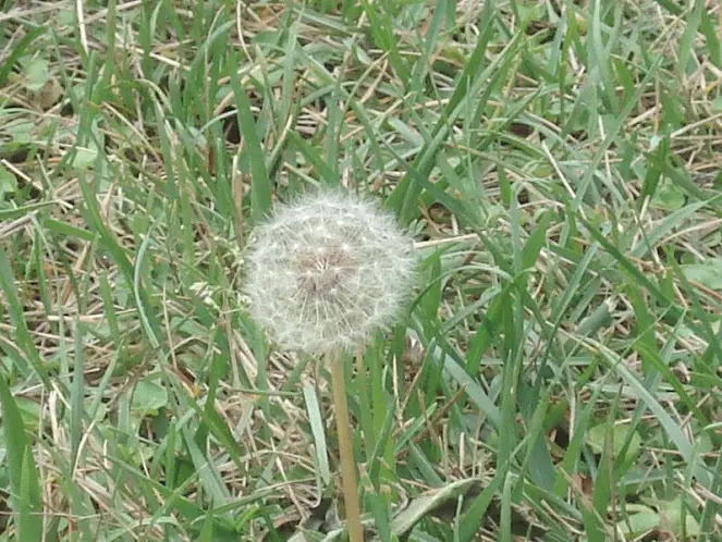
♢ 晩春のたんぽぽ・The Dandelion in the Late Spring
"Dandelion" is a seasonal word of March, but I photographed one till the end of April at last.
Because of my affection for the little plants, I like dandelions too. I like their fuzzy seeds, and they fling in the air on the wind.
In the morning, I only saw one dandelion. I have ever seen some others before this time, but after the rain and the strong wind did recently, all the seeds disappeared.
In this case, it's likely I would not see the full-sky dandelions' seeds fling this year.
However, I am pleasant. I am so satisfied even only for I saw the dandelions, and took photos for one.
In such a feeling, I prayed, for the seeds can fly in the air away distantly on the wind.
たんぽぽは弥生の季語ですが、この卯月の終わりのころ、私はやっと一つを撮られてきました。
小さな植物への愛着のため、私はたんぽぽをも好きです。特にその白い綿のような冠毛のこと。そんな種が風に乗って、満天飛んでいく光景をも好きです。
今朝、目に見えたのは一つしかありませんでした。この前にもいくつを見ることがあったのが、最近雨がよく降っていて、風も強かったし、みんなも瞬間に消えちゃいました。
こんな状況で、満天の飛んでいるたんぽぽを見られること、今年は無理なのそうですね。
そう言うけど、やっぱり喜びます。私やがて今年のたんぽぽを見られて、また写真を撮りましたの。これだけで、もう十分だと思います。
こんな心情で、私は祈りました。このたんぽぽの種たちが、風に乗られて、遠く遠く飛んでいられますように。